According to modern medical recommendations, every woman should undergo an annual preventive examination of a gynecologist.
At the reception the doctor interviews the woman’s health status, asking questions about the duration and nature of menstruation, the presence of gynecological diseases and disturbing symptoms. Then the doctor performs an examination of the patient on a gynecological chair, and takes the required tests.
Ultrasound of pelvic organs has an important role in the diagnosis of women’s diseases. Modern ultrasound machines can detect pathological changes in the female organs in the early stages.

Who and when a gynecological ultrasound is indicated
Ultrasound examination is recommended for women in the following cases:
- If during a visual examination, the gynecologist was unable to determine the cause of the complaints
- If a woman is planning a pregnancy, or an IUD (to rule out pathologies and contraindications)
- For preventive purposes, as part of an annual examination by a gynecologist.
- Many women’s diseases can be detected by ultrasound diagnosis at a very early stage, when symptoms are still absent.
What female abnormalities can be detected by ultrasound examination?
Some of the most common include:
1. endometriosis. This is a pathology in which the inner layer of the uterus (endometrium) grows outside of its cavity. On ultrasound, this can be seen by the presence of endometrial cells in the perineum, vagina or external genitalia. If the nidus also develops in the ovaries, heterogeneity of tissue and various nodular inclusions are visualized.
2. Uterine myoma. It is a benign neoplasm occurring in the myometrium – the muscular layer of the uterus. Exactly the ultrasound examination helps not only to determine the presence of the tumor, but also helps choose the treatment tactics. If myoma in the course of time does not increase in size, it will be enough just a doctor’s control. In other situations, the doctor prescribes conservative or surgical treatment.
3. Ovarian cysts. These are tumor-like formations filled with various contents. By gynecological ultrasound in this case, the presence of the cyst, its location and structure is determined.
4. Endometrial polyps. They are formations of benign nature of the endometrium of the uterus. On ultrasound examination, the inner mucous membrane of the organ looks dark, and polyps on its background are bright, light and with a clear contour.
A doctor cannot see polyps in the uterine cavity during a gynecological examination. If a woman is not bothered by anything, the abnormality can only be detected by ultrasound. Polyps are dangerous formations and require treatment.
5. Uterine adhesions. It is impossible to recognize adhesions with ultrasound. However, you can notice indirect signs of this pathology. Therefore, pelvic ultrasound examination should be included in the annual examination plan.
Important: Ultrasound of the female organs in most cases is most informative in the first phase of the cycle, ie, 5-7 days from the menstruation. But with low efficiency of the study appointed repeat procedure, as some pathology is better seen at the most pronounced endometrium – the 22-27 day of the cycle.
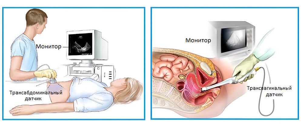
Types of Ultrasound in Gynecology
There are the following types of gynecological ultrasound:
transvaginal ultrasound. Examination is performed by inserting a special ultrasound transducer of anatomical shape into the vagina. With this method, the doctor can examine all of the female genital organs.
Transabdominal (or simply abdominal) ultrasound. It is performed through the front wall of the abdomen.